


Posted inDiagrams
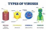
Structure Of Viruses Study
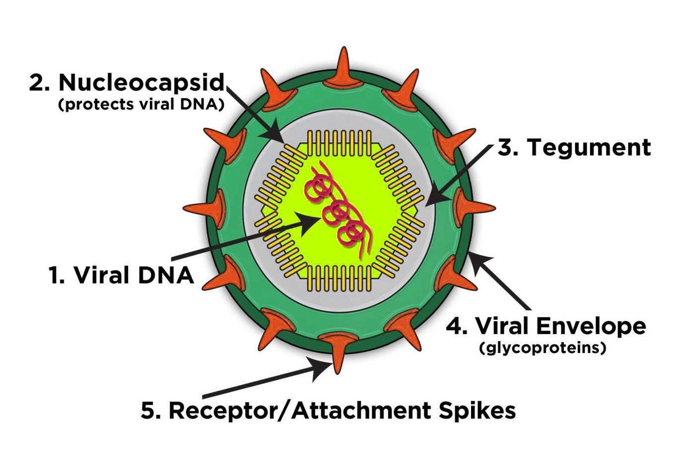
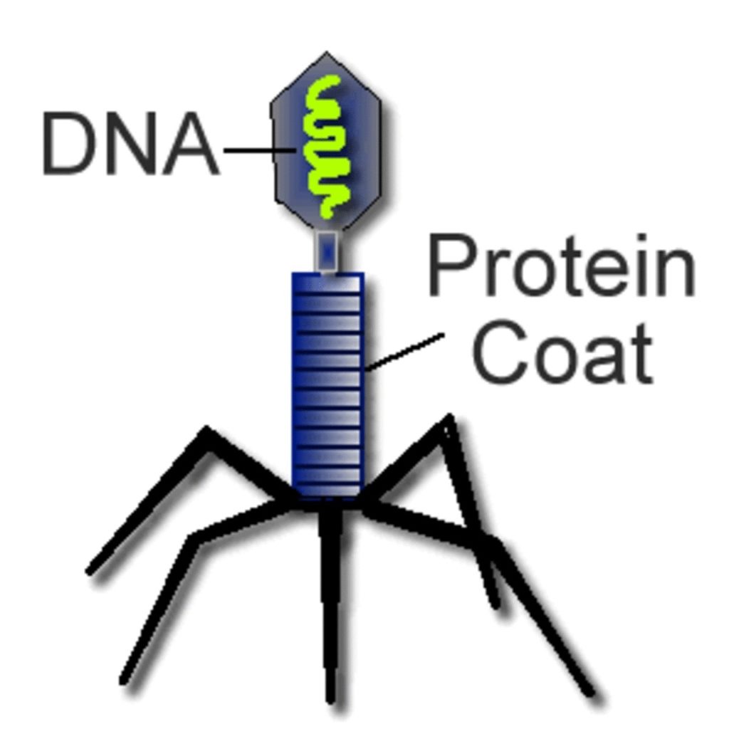
Structure of Viruses Viruses are biological structures that have a nucleic acid genome surrounded by protein and lipids. They are much smaller than bacteria and consist of a single- or…